Crowning a quest into a very well-guarded secret: Structure of the kinetochore corona finally revealed
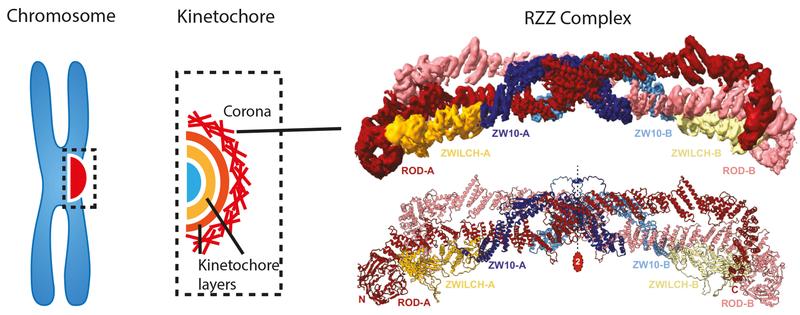
During cell division the 23 chromosomes must be first copied and later delivered to two newly forming daughter cells. At least in healthy cells, the result is astonishingly flawless, and no chromosome is ever lost. A multilayered protein structure called the kinetochore executes the chromosome delivery program. In a highly interdisciplinary collaborative tour-de-force, the groups of Andrea Musacchio and Stefan Raunser at the Max Planck Institute of Molecular Physiology studied the outermost layer of this structure, the kinetochore corona. They revealed the structural organization of the corona’s main building block, the RZZ complex, and deciphered the mechanism of corona assembly.
During cell division in a mother cell, the 23 chromosomes that carry the human genome must be first copied and later delivered to two newly forming daughter cells. At least in healthy cells, the result is astonishingly flawless, and no chromosome is ever lost. Not so in malignant cells, where rampant chromosome segregation errors generate a continuous flux of new genetic variants that support metastatic growth and resistance to chemotherapy. A multilayered protein structure called the kinetochore executes the chromosome delivery program. In a highly interdisciplinary collaborative tour-de-force, the groups of Andrea Musacchio and Stefan Raunser at the Max Planck Institute of Molecular Physiology studied the outermost layer of this structure, the kinetochore corona. With the help of single particle cryo electron microscopy and protein reconstitution they revealed the structural organization of the corona’s main building block, the RZZ complex, and deciphered the mechanism of corona assembly. Their results illuminate the molecular foundations of genome inheritance through the generations.
Cell division builds our bodies, supplying all cells in our tissues and organs, from the skin to the intestine, from the blood to the brain. It not only allows these organs to grow, but also to regenerate with fresh cells when required. Cell division starts with the replication of chromosomes, the carriers of the three billion nucleotides of the human genome. The replicated chromosomes are then distributed to the daughter cells in a process named mitosis. During mitosis, a network of thread-like structures named the mitotic spindle initially captures the chromosomes. After positioning them in a highly choreographed process, the spindle separates the chromosomes in opposite direction, so that when two cells form out of one, each inherits an exact copy of the genome. Even the smallest errors in this process will have dire physiological consequences.
A multilayered challenge
The kinetochore is the point of contact of chromosomes with the spindle, and is therefore crucially involved in the process of chromosome alignment and partition. It is a complicated multilayered protein complex. “Understanding kinetochores is a tremendous challenge, as they consist of several layers, each made of many interacting building blocks” says Musacchio. “The outermost layer, the corona, has retained some of the most interesting secrets of the kinetochore. Its assembly is particularly interesting, since the complex has a brief lifetime that ends right before the critical steps of chromosome alignment and segregation.“
In a series of previous studies, Musacchio’s laboratory made fundamental inroads into the structure and function of the different layers of kinetochores and how they connect chromosomes to microtubules. To gain this knowledge, the group adopted a reductionist approach named biochemical reconstitution. They produced the individual components of the protein networks outside the cell, in a test tube. They then reassembled them piece by piece to form an almost complete kinetochore that they could study in isolation, in a controlled and simplified environment that contrasts with the enormously complex, buzzing interior of a cell.
Applying the same strategy, the skilled team of two postdocs, Tobias Raisch and Giuseppe Ciossani, two PhD students, Ennio d’Amico and Verena Cmentowski, and other co-workers has now been able to rebuild the kinetochore corona. They showed that only two components are sufficient for that: the ROD-Zwilch-ZW10 (RZZ) protein complex and the protein Spindly, which plays an essential role in the interaction of the kinetochore with the microtubules. The corona assembles exclusively on kinetochores, and the mechanisms that limit its growth to these structures had remained a crucial unresolved question. By reconstituting the process in vitro, the scientists were able to identify an enzyme, the kinase MPS1, as the essential catalyst of RZZ corona assembly at the kinetochore.
One step closer to the crown
Electron microscopy (EM) has accompanied the study of kinetochores since the 1960’s, but it wasn’t until recently, that burgeoning methodological developments made this technique able to visualize the building blocks at the atomic scale. “In 2017, we generated the first ever 3D structural model of the RZZ complex by cryo-EM”, says Raunser. “However, at the 1 nm resolution of this initial model, it was impossible to observe the finest molecular details responsible for biological function.”
The new structural analysis improved the resolution to the point that atomic details emerged, finally explaining how interactions of RZZ components with themselves and with Spindly promote corona assembly into a large polymer that surrounds the kinetochore. “Our work crowns a succession of previous studies on the kinetochore corona, now providing us with a framework to understand the critical moment of cell division when the attachment of chromosomes to microtubules becomes essentially irreversible” concludes Musacchio. The team’s future studies will try to integrate the corona into reconstituted kinetochores, moving a new important step towards the reconstitution of chromosome segregation in vitro, a goal of extraordinary ambition that will shed light on the basis of a most fundamental process of life.
Wissenschaftlicher Ansprechpartner:
Prof. Dr. Andrea Musacchio
Max-Planck-Institut für molekulare Physiologie
Mechanistische Zellbiologie
Tel.: +49 231 133 2101
E-Mail: andrea.musacchio@mpi-dortmund.mpg.de
Originalpublikation:
Raisch T, Ciossani G, d'Amico E, Cmentowski V, Carmignani S, Maffini S, Merino F, Wohlgemuth S, Vetter IR, Raunser S, Musacchio A. Structure of the RZZ complex and molecular basis of Spindly-driven corona assembly at human kinetochores. EMBO J. 2022 Apr 4:e110411. doi: 10.15252/embj.2021110411
Weitere Informationen:
https://www.mpi-dortmund.mpg.de/news/crown-kinetochore
Ähnliche Pressemitteilungen im idw


