University of Applied Sciences Hof and SANA Klinikum Hof: How 3D Printers and a Storage Container Can Become Lifesavers
Hof, Germany – An innovative training opportunity for aspiring doctors has become a reality thanks to a partnership between the University of Applied Sciences Hof and SANA Klinikum Hof. Through this collaboration, young doctors can now practice minimally invasive procedures for treating vascular occlusions on a life-like model—an innovation poised to revolutionize medical education. Previously, such training was only possible under the guidance of highly experienced colleagues on patients or animal models. This cost-effective solution is expected to become available to other medical universities and training institutions in the future.
The MakerSpace at the University of Applied Sciences Hof is a state-of-the-art workshop equipped with high-tech machines, tools, and software, enabling the realization of technical ideas and prototype development. These impressive resources caught the attention of Mohammed Misbahuddin-Leis, a resident in diagnostic and interventional radiology, as he hurried through downtown Hof with his idea. René Göhring, Technical Director at MakerSpace, recalls with a smile: “One afternoon, a young resident in a lab coat came to us and asked if we could build something for him. It seemed as though he had dashed over between surgeries. You could sense the urgency of his project.”
Improved Training Opportunities for Aspiring Radiologists
What began as an idea has now become a success. The "MANTA 3.4" – the "Medical Angiography Training Phantom for Doctors" – is a model that allows medical trainees to practice handling catheters and closure techniques in a realistic environment without immediately operating on patients. Prof. Dr. Boris Radeleff, Chief of Diagnostic and Interventional Radiology at SANA Klinikum Hof and adjunct professor at Heidelberg University Hospital, describes the progress: “Vascular occlusions often need to be resolved within minutes. Precision and speed are crucial. Until now, practical training opportunities were extremely limited. Trainees had to practice either under the supervision of experienced colleagues directly on patients—a risky approach and limited by the rarity of certain bleeding cases—or on animal models, which extended the learning curve. With our new model, we can significantly enhance training for our budding radiologists, ultimately improving patient care.”
Developing a Training Phantom
Previously, the only high-functioning yet expensive phantom available was the AngioTrainer, developed by Charité in Berlin. Due to its cost, broad training of medical staff, particularly in countries with limited medical resources, is either severely restricted or only possible at a few elite centers. "The fact that we can now implement this together with the University of Applied Sciences Hof is a significant advancement," says Dr. Radeleff. "Our goal was to create a cost-effective training phantom that could be manufactured for training purposes at universities and training centers with limited resources," explains Prof. Dr. Anke Müller, Professor of Manufacturing Technologies and Head of the Startup Lab at the University of Applied Sciences Hof.
Preparing for Emergencies
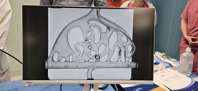
In the MakerSpace, models of human vascular structures were created using 3D printers, with an integrated variable pump system. This system simulates human arterial blood pressure, flow rates, and physiological conditions encountered during real procedures. “After several prototypes, we developed a version that could accurately replicate a transparent, flexible vascular system,” reports Prof. Dr. Thomas Rausch, a key contributor to the project. This enables trainees to safely practice handling instruments such as catheters and guidewires and using coils (spirals) for closure. Prof. Radeleff adds: “Our MANTA phantom prepares medical professionals for emergencies and enhances their responsiveness and precision during urgent medical interventions. Training scenarios such as acute vascular occlusions or arterial bleeding allow interventional radiologists to act faster and more accurately.”
Cost-Effective and Easy to Produce
The simplicity and affordability of the model, originally developed using a 3D printer and a storage container, are key advantages that increase the accessibility of training for a broader range of medical students. “The material costs for the model are approximately €250. Additionally, all components can be produced with equipment commonly available in university environments,” says Prof. Dr. Anke Müller. Plans include making the technology available as an open-source guide for other training institutions and university hospitals, in line with the maker movement.
The future of medical training looks promising: The model will continue to evolve, incorporating specific training modules for various radiological sub-disciplines. “This innovation sets a new standard in medical education, ultimately benefiting our patients,” emphasizes Prof. Radeleff. Requests have already been made for models for urology.
Wissenschaftlicher Ansprechpartner:
Prof. Dr.-Ing. Anke Müller
+49 9281 409 - 4680
anke.mueller(at)hof-university.de
Ähnliche Pressemitteilungen im idw


